Lipid Membrane Phase Separation and Protein Sorting
How do lipids organize proteins within cell membranes, and can membrane composition control biological function?
Cell membranes are not homogeneous fluids. Their complex mixtures of lipids and cholesterol can give rise to regions with distinct physical properties, including liquid-ordered (Lo) and liquid-disordered (Ld) phases. This lateral organization can strongly influence where membrane proteins accumulate, how they interact and ultimately how they function.
At BendixLab, we investigate how lipid phase separation, membrane curvature and molecular interactions govern the spatial organization of membrane proteins. Understanding these physical mechanisms is essential for explaining cellular signaling, membrane trafficking, viral assembly and other processes that depend on the organization of proteins within biological membranes.
Quantifying Protein Sorting in Lipid Membranes
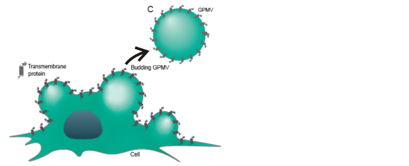
Using fluorescence microscopy, optical tweezers and giant plasma membrane vesicles (GPMVs) derived from living cells, we have developed quantitative approaches to investigate how proteins partition between different lipid environments.
Our studies have demonstrated that membrane proteins exhibit distinct preferences for lipid phases and membrane geometries. In particular, we showed that the influenza membrane protein neuraminidase preferentially partitions into liquid-disordered membrane regions, whereas the membrane repair protein annexin A5 exhibits strong curvature-dependent enrichment. These findings illustrate how lipid composition and membrane geometry provide complementary physical mechanisms for organizing proteins.
Engineering Membrane Composition Using Thermoplasmonic Fusion
A major challenge in membrane biophysics is studying proteins in well-defined lipid environments while preserving their native membrane orientation and structure.
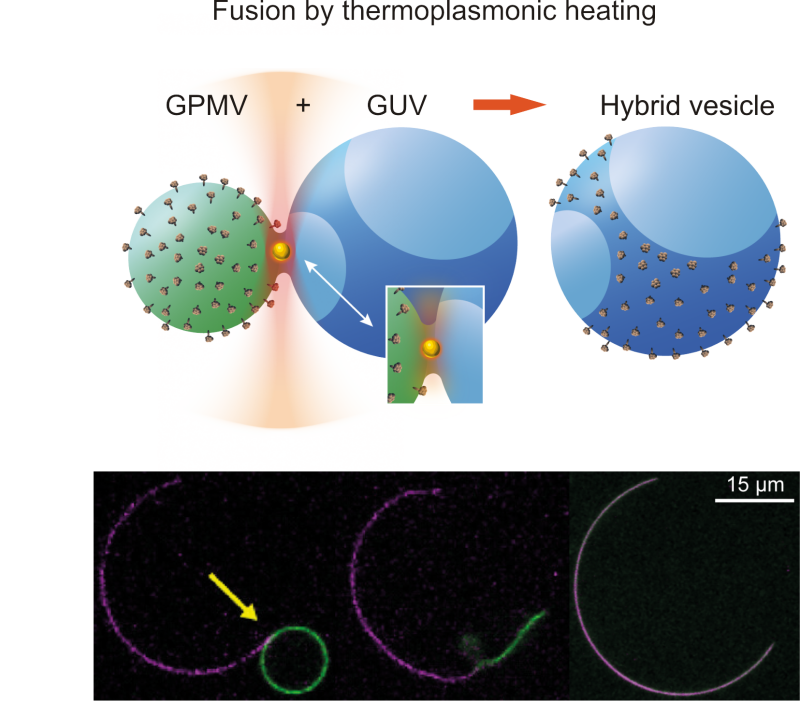
To overcome this, we developed a method combining optical trapping and thermoplasmonic membrane fusion to transfer membrane proteins from cell-derived vesicles into phase-separated artificial lipid membranes.
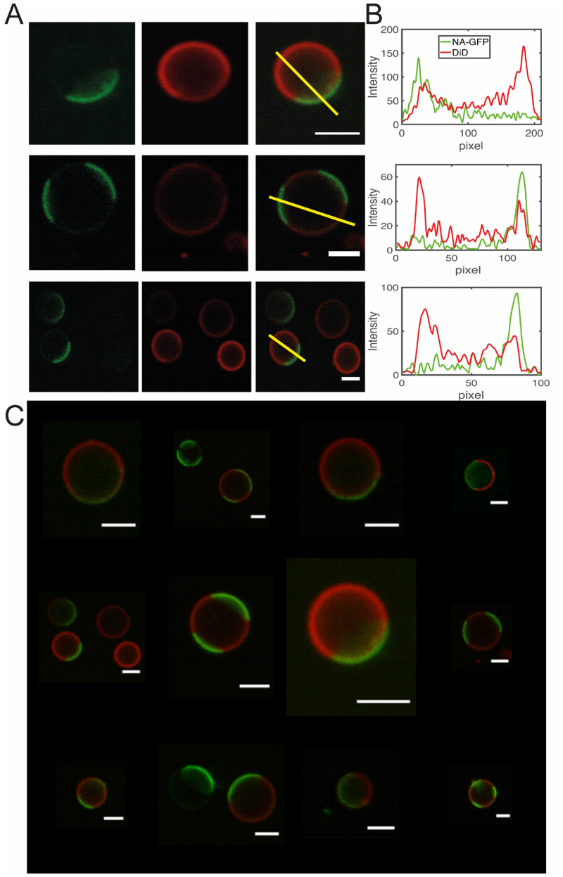
By selectively fusing GPMVs with giant unilamellar vesicles (GUVs), we create hybrid membranes that combine biologically expressed proteins with controlled lipid compositions. This allows us to visualize and quantify protein partitioning between liquid-ordered and liquid-disordered domains without the need for protein purification.
Using this approach, we demonstrated that the influenza A virus spike proteins hemagglutinin (HA) and neuraminidase (NA) preferentially partition into liquid-disordered lipid phases. This finding provides new insight into the physical principles underlying viral membrane organization and raises important questions about the role of cholesterol-rich membrane domains during viral assembly.
Connecting Lipid Organization to Biological Function
Our broader goal is to understand how the physical properties of lipid membranes regulate protein localization and activity. We are particularly interested in how lipid packing, cholesterol content, phase separation and membrane curvature interact to control the organization and function of transmembrane proteins, including G-protein-coupled receptors (GPCRs).
By combining controlled membrane engineering with quantitative fluorescence imaging and mechanical manipulation, we aim to reveal how membrane composition and geometry influence biological signaling at the nanoscale.
Selected Publications
-
Moreno-Pescador et al. (2023). Thermoplasmonic Vesicle Fusion Reveals Membrane Phase Segregation of Influenza Spike Proteins. Nano Letters 23, 3377–3384.
DOI: https://doi.org/10.1021/acs.nanolett.3c00371 -
Moreno-Pescador et al. (2019). Curvature- and Phase-Induced Protein Sorting Quantified in Transfected Cell-Derived Giant Vesicles. ACS Nano 13, 6689–6701.
DOI: https://doi.org/10.1021/acsnano.9b01052 -
Andersen et al. (2014). Nanoscale Phase Behavior on Flat and Curved Membranes. Nanotechnology 25, 505101.
DOI: https://doi.org/10.1088/0957-4484/25/50/505101 - Andersen et al. (2014). Local and transient permeation events are associated with local melting of giant liposomes. Soft Matter 10 (24), 4268-4274. DOI: https://doi.org/10.1039/c4sm00410h